“My stained glass work is a fusion of science and art.”
Joel Kowit
Artist Statement: The Cellular Universe Through Stained Glass
What does it mean to fuse science and art? How does this fusion contribute to art, to science, to society, and to the viewer?
BioArt takes many forms: images of cells, tissues, organisms, taken with electron or light microscopes, sometimes landscape-like, sometimes abstract patterns of balance and color; bacterial cultures forming unpredictable patterns on agar plates; sculptures of imaginary creatures produced by genetic engineering in the minds of the artist, and much more. Some of these art forms pose ethical questions about the science and its implications for society. Others are more about the beauty, harmony of the work, and the emotions the work evokes in the viewer.
Where does my artwork fit in this picture? Stained glass is a thousand-year old technology, historically used in church windows to inspire awe and reverence for biblical characters and for God. My work focuses on the cellular, rather than the celestial universe: biomolecules and cellular structures - the characters of life. The medium of stained glass parallels the microscope, which relies on the transparent surface of the cell to visualize stained internal structures. I hope that the artistic qualities alone justify these works – color, harmony, energy, balance, movement, and light – especially light since stained glass is unique in how the changing light of weather, season, and time-of-day are integral to the artwork itself.
But these artworks are also iconic images of science. Each piece has a story, often related to my own experiences as a scientist - something I worked on in the lab, or taught in the classroom, or lectured on in my career. Several are inspired by work associated with Nobel Prizes in chemistry, medicine or physiology; some are associated with important human diseases.
Images of science, they are scientifically accurate within the context of the art form. One group of these stained-glass pieces show structures of proteins, e.g., HIV Protease (“Inhibition”), Potassium Channel (“Passage”), Beta-amyloid Fibril (“Loss”). These protein structures have been solved, to atomic resolution, by x-ray diffraction (or similar techniques.) The atomic coordinates are deposited in a freely accessible national database, and all my protein artworks use these PDB/protein database files to create models on which are based the designs for the stained-glass work. Similarly, the cellular structures – Organ of Corti (“Vibration”), Retina (“Vision”), 26S Proteasome (“Degradation”), Muscle Sarcomere ("Contract"), – are based on our knowledge of the actual structures and how they function.
The scientific accuracy of the artwork is important. Scientific honesty is fundamental to good science and these artworks can facilitate learning science – a form of knowledge visualization. I hope that non-scientists, or budding scientists, viewing these pieces, will allow their curiosity to entice them along the path to science and to a greater understanding of the fundamental question – “How does life work?”
Stained Glass Exhibits
CURRENT:
Solo Exhibition, Harvard Medical School, NRB/New Research Building, 77 Avenue Louis Pasteur, Opening: July 3, 2019 continuing through Spring, 2021. Opening reception and artist talk, July 24, 2019, 4-6 pm “The Cellular Universe Through Stained Glass” (11 pieces) https://libcal.countway.harvard.edu/event/5541058
https://www.facebook.com/martinconferencecenter/
PAST:
Solo Exhibition, Cary Memorial Library, Lexington, MA (March 6-April 30, 2018) (8 stained glass pieces plus 5 framed pieces showing photo collages of the process of stained glass.
Solo Exhibition, LabCentral, Kendall Square, Cambridge (June 26-August 23, 2017) (13 stained glass pieces, plus 22 framed pieces showing preliminary drawings, sketches, and photo collages of the process of stained glass.)
Gallery Blink, Lexington, MA January, 2017 (2 pieces)
Pieces sold:
Vibration (Organ of Corti)
Decibel Therapeutics, Boston, MA
Loss (Beta-Amyloid Fibril)
LabCentral, Kendall Square, Cambridge, MA
Degradation (26S Proteasome)
Dr. Alfred Goldberg, Professor of Cell Biology, Harvard Medical School
Migration (Leukocyte Extravasation)
Dr. Rodger McEver, Vice President of Research, Oklahoma Medical Research Foundation
Inflammasome Series (I)
Dr. Peter Libby, former Chief of Cardiovascular Medicine, Brigham & Women’s Hospital; Mallinckrodt Professor of Medicine, Harvard Medical School
Inflammasome Series (2)
Dr. Paul Ridker, Director, Center for Cardiovascular Disease Prevention, Brigham and Women’s Hospital, and Eugene Braunwald Professor of Medicine, Harvard Medical School
Inflammasome Series (3)
Dr. Eicke Latz, Institute Director, Institute of Innate Immunity, University of Bonn, Germany
Inflammasome Series (4)
Anonymous
Articles
Summer Newsletter, July, 2019, issue 82 (online only); RCSB-PDB [Protein Data Bank], “How Does Life Work? Creating Scientific Explanations in Stained Glass” by Joel Kowit https://cdn.rcsb.org/rcsb-pdb/general_information/news_publications/newsletters/2019q3/corner.html
Joel Kowit met les virus sous verre [“Joel Kowit puts viruses under glass” ] https://www.liberation.fr/sciences/2020/03/31/joel-kowit-met-les-virus-sous-verre_1782733
Profile: A Scientific Perspective on Art (written by Nidhi Sharma), Chemical Engineering Progress, February, 2020
Honors
“Cas9” - Cover Image, The CRISPR Journal, February, 2020
“Degradation” (26S Proteasome) - Cover Image, Biochemical Journal, 474(19) 2017
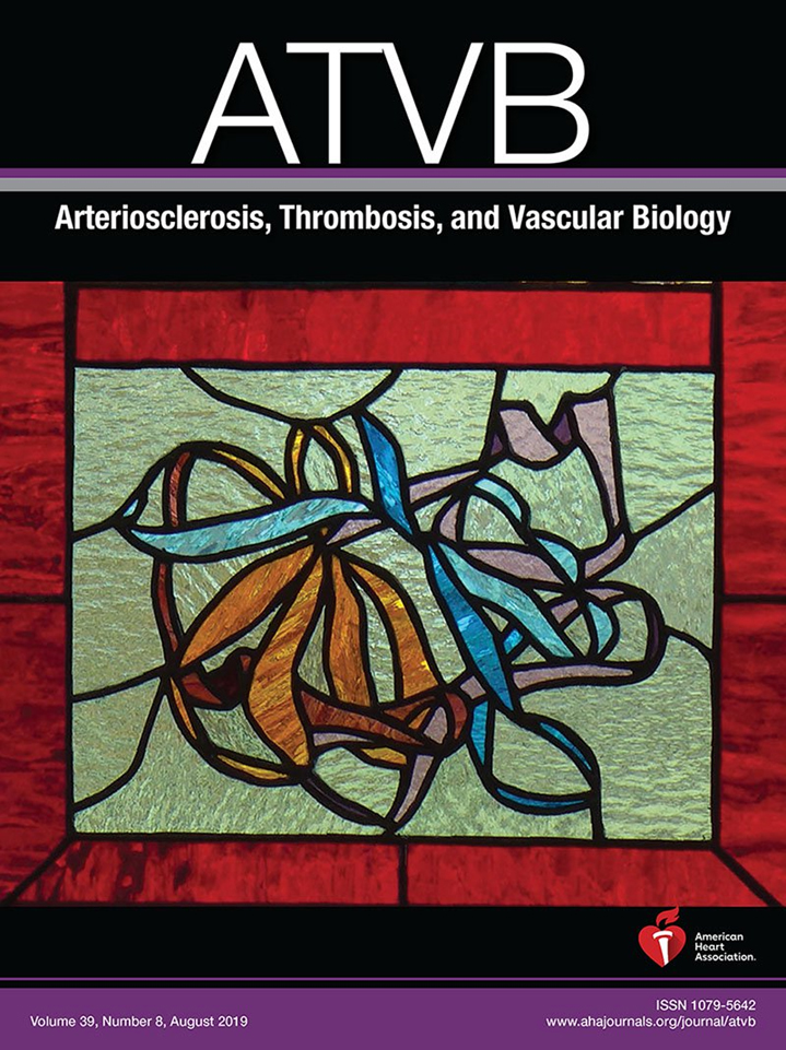
“Inflammasome” (Detail) - Cover Image, Arteriosclerosis, Thrombosis, and Vascular Biology, 2019, August; vol 39, #8
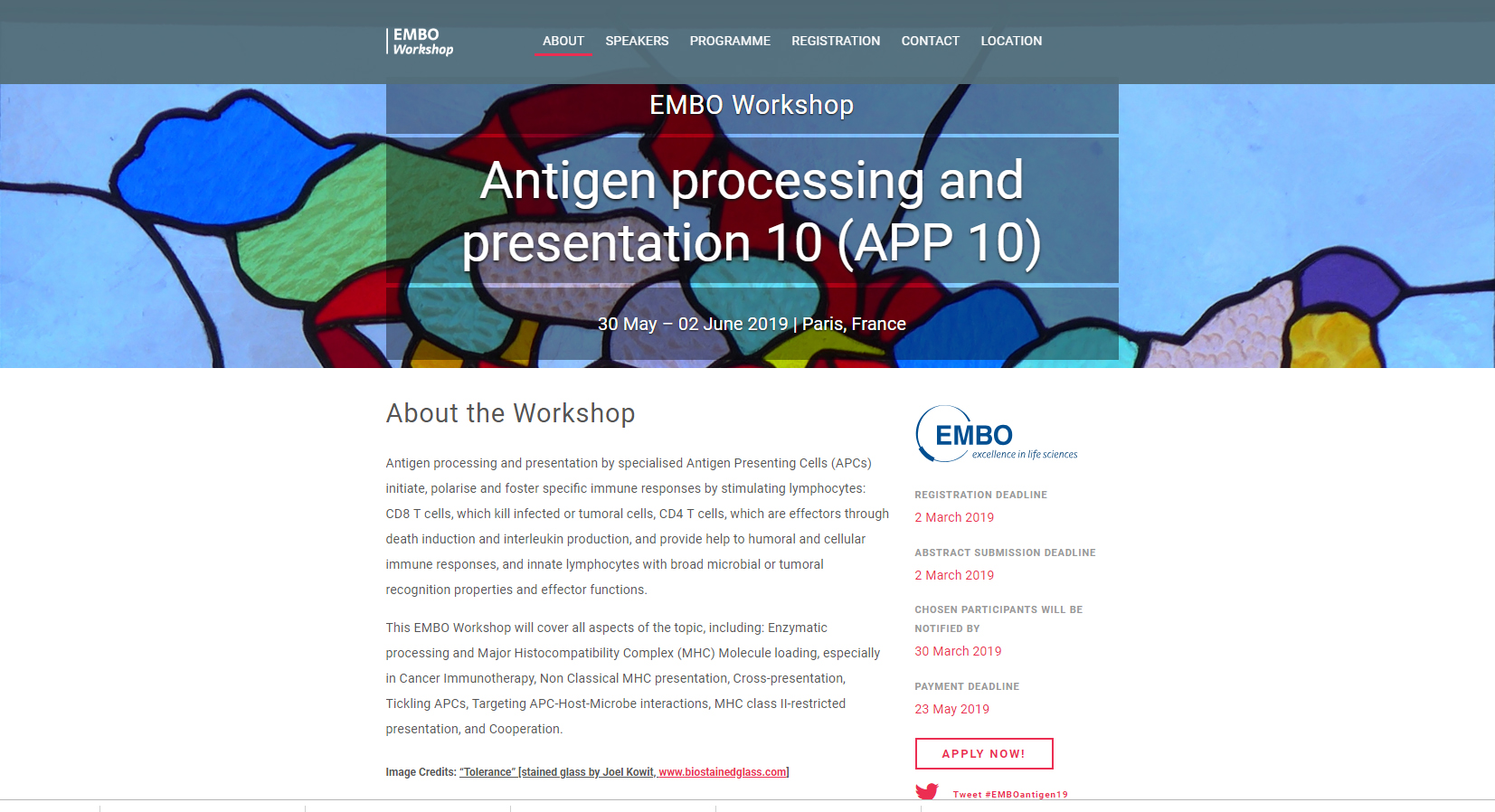
Tolerance” (MHC II/peptide) image chosen for website / EMBO Workshop, Paris, 2019
Breakthrough Artist of the Year Award, Council for the Arts, Lexington, MA for 2017 for stained glass work (First year for the award)
Scientific Journal Covers
Inflammasome (detail showing IL-1 beta panel)
Tolerance” (MHC II/peptide) image chosen for website / EMBO Workshop, Paris, 2019
Painting Exhibits
Solo Exhibition, Massachusetts State House, Boston, 2013
Solo Exhibition, Gallery 5, Emmanuel College, Boston
Solo Exhibition, Lincoln Library, Lincoln, MA, 2014
Solo Exhibition, Cary Memorial Library, Lexington, MA, 2013
Solo Exhibition, Cary Memorial Library, Lexington, MA 2009
Twitter stuff
https://twitter.com/PDBeurope/status/921670407257776128
https://twitter.com/search?q=joel%20kowit&src=typd


